Cut your overnight blots down to a single workday while boosting reproducibility
1 Why Western blotting still hurts
Despite its status as the gold-standard for validating protein expression, the classic workflow is dogged by:
|
Pain point |
Typical cause |
Impact |
|---|---|---|
|
Time-to-result |
Overnight antibody incubations plus long transfer runs |
≥ 16 h turnaround |
|
Transfer inefficiency & overheating |
Wet-tank transfers at 100 V for ≥ 1 h |
Patchy bands, distorted high-MW proteins |
|
Hands-on variability |
Manual blocking/wash cycles |
User-to-user CV > 20 % in band intensity |
|
Antibody cost |
Large incubation volumes, single use |
Primary Ab spend can exceed SGD 5 k/yr/lab |
|
Dynamic-range limits |
Film or narrow-range CCD imagers |
Weak bands lost; strong bands saturated |
2 Technology advances that shrink the workflow
|
Workflow segment |
Recent innovation |
Time saved |
Key benefit |
|---|---|---|---|
|
Gel run |
Pre-cast gradient or stain-free gels (Bio-Rad TGX Stain-Free, Bolt Bis-Tris) |
30–40 min |
No casting; fluorescence imaging of total protein for loading control |
|
Protein transfer |
Semi-dry rapid transfer – Trans-Blot Turbo (3 min) ; Dry transfer – iBlot 3 (≤ 7 min) |
≥ 50 min |
Less heat, consistent high-MW recovery |
|
Blocking / Ab incubations |
Automated processors (e.g., Bandmate, SNAP i.d) recycle antibodies and run unattended |
1–4 h |
Up to 90 % hands-on reduction; 3-fold Ab economy |
|
Detection |
Near-IR imagers (LI-COR Odyssey DLx) give 6-log dynamic range; capillary “blots” (Bio-Techne Jess) finish in ≈ 30 min |
≥ 1 h |
True quantitation, multiplex two targets at once |
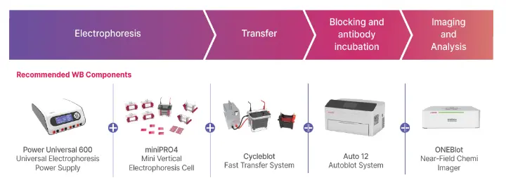
3 Putting it together: the integrated 5-hour workflow we distribute
By chaining the fastest option in each segment, a complete blot—including imaging and densitometry—fits comfortably inside a standard workday:
|
Step |
Module (generic) |
Runtime |
|---|---|---|
|
SDS-PAGE (4-gel mini tank, 200 V) |
45 min | |
|
Rapid semi-dry transfer (pel-tier cooled) |
20 min | |
|
Automated block → 1 h primary (37 °C) → 15 min secondary |
90 min | |
|
Near-field chemiluminescent or IR imaging + analysis |
5 min | |
|
Total bench-to-result |
≈ 4 h 45 min |
How it fixes the pain points
- 70 % faster than the legacy 16 h protocol.
- Uniform transfer mitigates “smiling” and overheating artefacts.
- Closed-loop incubations cut antibody use by up to 5-fold.
- 6-log detection range means both faint PTMs and abundant housekeeping bands sit in the linear zone.
4 Technical tips & troubleshooting
- Sample prep: Keep lysates ≤ 4 °C; spin 16 000 g, 15 min to remove debris.
- Gel % logic: 4–20 % gradient for unknowns; 8 % for 70-250 kDa; 12 % for < 30 kDa.
- Voltage ramp: Start gels at 80 V until the dye front stacks, then jump to 200 V to finish in < 45 min.
- Membrane choice: PVDF for hydrophobic or low-MW proteins; rinse briefly in methanol then transfer buffer.
- Rapid transfer packs: Pre-wet membrane and stack only once to avoid air bubbles—major cause of uneven bands.
- Automated processor set-up: Use 3–5 mL/block (mini blot) and enable “antibody recovery” to reuse primaries 3 ×.
- Imager QA: Run the built-in glow-ladder once per quarter; check that R² > 0.99 across the intensity range.
- Quantitation best practice: Always normalise to total-protein stain (stain-free or REVERT total-protein IR stain) instead of housekeeping genes for higher accuracy.
References
- Bio-Rad Laboratories. The Complete Guide to Stain-Free Western Blotting (2024).
- Thermo Fisher Scientific. iBlot 3 Dry Blotting System Manual (2024).
- Bio-Rad Laboratories. Trans-Blot Turbo Transfer System product page (accessed 24 May 2025).
- Thermo Fisher Scientific. Bandmate Automated Western Blot Processor specifications (accessed 24 May 2025).
- LI-COR Bio. Odyssey Imager Family overview (accessed 24 May 2025).
- Bio-Techne. Jess Automated Western Blot System – Benefits and Workflow (2025).
- Tanabe L. The Ultimate Quick Tips & Tricks Guide to Western Blots. Biocompare, 9 May 2024.