
1. A virus meets a variable defense
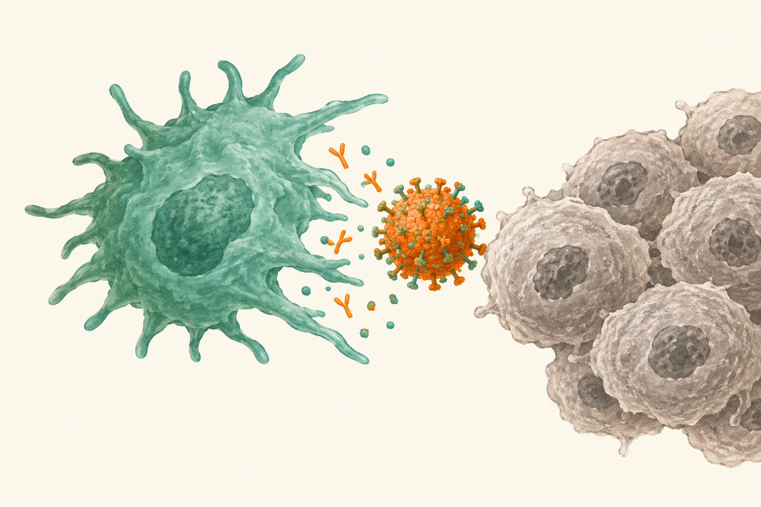
Oncolytic virus M1 can suppress some mouse tumors, yet others barely respond. What inside a tumor might stop the virus before it helps recruit an immune attack? Liang and colleagues investigated tumor-associated macrophages—immune cells that can fight tumors but also restrict viruses (1). A review describes this antiviral and antitumor tension as a recurring challenge for oncolytic-virus therapy (2).
2. Comparing tumors and changing macrophage numbers
The researchers treated mice bearing one of four subcutaneous tumor models: colorectal MC38, pancreatic KPC1199, prostate RM1, or melanoma B16F10. They compared tumor growth with controls, measured macrophage populations by cell-marker analysis, and counted viral RNA in tumors over time. In further experiments, they reduced macrophages using either clodronate-containing liposomes or antibodies against the macrophage-supporting receptor CSF1R. Immune-cell profiling, tumor-growth measurements, and survival tracking tested what changed; a separate experiment depleted CD8-positive T cells to test whether those cells were needed (1).
3. Where macrophages were abundant, response was weaker
Two models met the authors’ response threshold—a treated-to-control relative tumor-growth ratio of 50% or less—while MC38 and KPC1199 exceeded it. Across the four models, macrophage infiltration tracked that ratio closely (R²=0.9618; p=0.0193). This suggests macrophage abundance may help explain variable response, but four model-level comparisons cannot establish a predictive marker for patients. The association was also strong for M1-like macrophages (R²=0.9345); the M2-like association did not reach statistical significance (1).
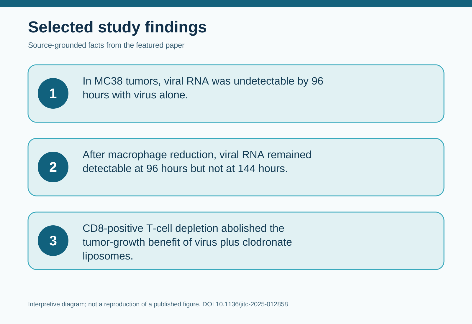
In MC38 tumors, viral RNA became undetectable by 96 hours with virus alone. After macrophage reduction, it remained detectable at 96 hours but not at 144 hours. The authors infer that macrophages restrict intratumoral virus. More persistent RNA supports that account, although RNA counts do not themselves prove infectious-virus replication or identify how macrophages removed it (1).
4. A stronger response needed CD8-positive cells
In MC38 tumors, the combination increased the proportion of granzyme-B-positive CD8-positive T cells relative to each other group (p<0.05). That finding is consistent with a stronger cytotoxic immune response, but a cell-marker proportion alone cannot establish which cells killed tumor cells. Neither virus nor either macrophage-reduction approach alone significantly restrained MC38 growth; both combinations slowed growth and extended mouse survival. Removing CD8-positive T cells abolished the growth benefit of virus plus clodronate-containing liposomes. Similar combination benefits appeared in the other three models, including the initially weakly responsive pancreatic model (1).
5. Limitations and uncertainty
These were implanted mouse tumors, not tests of treatment benefit or safety in people. Macrophage-reduction methods also affected cells outside tumors, including blood macrophages or monocytes, so their effects cannot be assigned solely to tumor macrophages. The M1-like subset was associated with resistance but was not selectively removed; the proposed clearance mechanism remains an inference requiring direct tests. The study did not predetermine sample sizes statistically, and its strongest correlation spans only four tumor models (1).
6. Takeaway
In these mouse models, reducing macrophages gave the virus more time in tumors and made its growth-control benefit depend on CD8-positive T cells. The practical research question is whether a more selective approach can preserve useful macrophage functions while improving viral therapy (1).
7. References
(1) Liang X, Li J, Chen J, Chen C, Li H, Yan C, Guo C, Han Y, Liu W, Sai K, Lin Y, Yan G, Zhu W, Liu Y. TAMs-mediated resistance to oncolytic virus M1 in solid tumors. Journal for immunotherapy of cancer. 2025;13(11):e012858. doi:10.1136/jitc-2025-012858.
(2) Lecoultre M, Walker PR, El Helali A. Oncolytic virus and tumor-associated macrophage interactions in cancer immunotherapy. Clinical and Experimental Medicine. 2024. doi:10.1007/s10238-024-01443-8.
8. Topics
#OncolyticViruses #TumorMacrophages #CancerImmunology #MouseModels