
1. The puzzle: shared signals, different cancers
Can one small set of DNA methylation markers distinguish tumors from nearby noncancerous tissue across eight cancer types? Tumors often alter methylation, but a marker useful in one organ may perform poorly in another. Neefs and colleagues tested whether three selected markers could work together across breast, colorectal, lung, prostate, pancreatic, liver, esophageal, and head-and-neck cancers (1).
A separate review notes that bulk methylation measurements can conceal rare cellular patterns; this study did not examine individual cells (2).
2. How was the assay tested?
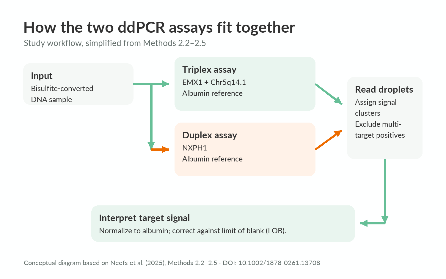
The researchers selected three methylation targets after screening candidate sites and testing whether primers distinguished cancer-cell-line DNA from healthy blood DNA. They built two droplet digital PCR reactions: one measured two targets plus a DNA reference, and the other measured the third target plus the reference. Dividing DNA into droplets let them count target-positive reactions (1).
They tested retrospectively collected, fresh-frozen samples: 103 tumors and 109 normal-adjacent tissues across the eight cancer types. Pathology review confirmed tissue type and estimated tumor-cell content. The team used five-fold cross-validation to assess classification within this tissue dataset; it was not a blood-based screening study (1).
3. What did the individual markers detect?
Two targets showed statistically significant tumor-versus-normal-adjacent methylation differences in all eight tissue types; the remaining target did so in seven, but not colorectal tissue. This suggests shared signals are measurable, not that every marker works equally well for every cancer. Across all samples, individual-target sensitivities were 82.5%, 76.6%, and 74.3%, respectively (1).
The laboratory detection limits were 0.84%, 1.45%, and 1.75% methylation for the three targets. These are measurements under assay-testing conditions, not the smallest tumors detectable in people. Tumor methylation also varied widely, so a weak signal in one sample cannot be assumed to mean an early-stage cancer (1).
4. What changed when targets were combined?
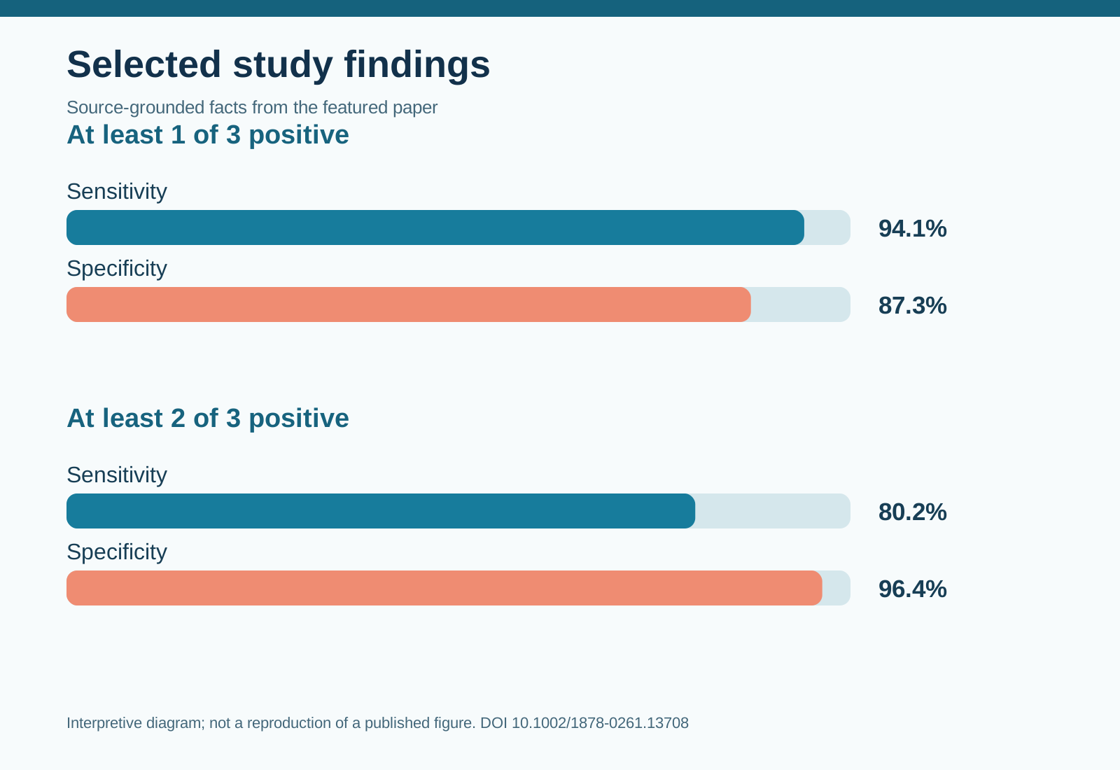
With a positive result defined as methylation at any one of the three targets, sensitivity was 94.1% and specificity was 87.3%. Requiring at least two positive targets shifted sensitivity down to 80.2% and specificity up to 96.4%. In plain language, the stricter rule missed more tumors in this dataset but classified more normal-adjacent samples correctly; neither result establishes performance in a screening population (1).
The combined targets had a cross-validated area under the curve of 0.948, summarizing discrimination across thresholds in these samples. The authors infer that combining targets supports multi-cancer detection. More cautiously, the result supports further testing of tumor-versus-adjacent-tissue classification, not a ready-to-use clinical test (1).
5. Limitations and uncertainty
Normal-adjacent tissue is not the same comparison group as blood from people without cancer. Some cancer subgroups were small: esophageal cancer had only five normal-adjacent samples, below the authors’ sample-size target of seven. Cluster assignment involved manual decisions, which may complicate standardization. The assay did not identify a tumor’s tissue of origin, and its performance in blood-derived cell-free DNA or routinely processed tissue remains untested (1).
6. Takeaway
Three methylation targets distinguished tumors from nearby tissue with a clear sensitivity–specificity trade-off. The useful next question is whether that trade-off holds in independent samples and in the specimen types a clinical test would actually use (1).
7. References
(1) Neefs I, De Meulenaere N, Vanpoucke T, Vandenhoeck J, Peeters D, Peeters M, Van Camp G, Op de Beeck K. Simultaneous detection of eight cancer types using a multiplex droplet digital PCR assay. Molecular oncology. 2025;19(1):188–203. doi:10.1002/1878-0261.13708.
(2) Kim IS. Single-cell DNA methylation profiling: Technologies, computation, and applications in precision oncology. Molecular oncology. 2026. doi:10.1002/1878-0261.70303.
8. Topics
#DNAMethylation #CancerBiology #MolecularDiagnostics #Biomarkers